
Veterinary Continuing Education (14 RACE-approved hours*).
American speakers - In France. Très bon!
In 2022 we're fortunate to have 2 amazing speakers

Howard Seim III, DVM, DACVS, Professor of Surgery, College of Veterinary Medicine, Colorado State University. Dr. Seim graduated from Washington State University, completed an internship in Saskatoon, Saskatchewan Canada, and a surgical residency at the Animal Medical Center in New York City. He obtained Diplomate status in the American College of Veterinary Surgeons in 1983. He is currently Professor of Surgery at Colorado State University and has been awarded the Merck AGVET Award for Creative Teaching, the CSU Award for Instructional Innovation and selected as the North American Veterinary Community’s Small Animal Speaker of the Year in 2009. Dr. Seim is founder of VideoVet, a Veterinary Surgery Continuing Education video series.
Generic lecture description
These sessions will consist of a variety of practical soft tissue surgery techniques that most veterinarians can perform in their practice. Video segments of clinical case material, carefully edited to form a real-life experience, will be used as a means of delivering the surgical lectures. An advantage of this lecture style is that participants are able to see the case actually operated on during the lecture.
Surgical management of gall bladder mucocele
Surgical management of gall bladder mucocele varies somewhat depending on the stage of gall bladder mucocele presentation; i.e., early presentation, late presentation, or presentation after gall bladder rupture. Video examples of each presentation will be used to illustrate the impact each stage of presentation has on the difficulty of surgical manipulation during cholecystectomy.
Visceral organ biopsy
This seminar will illustrate a variety of biopsy techniques of abdominal visceral organs. Discussion will include biopsy techniques used to acquire diagnostic samples of liver, pancreas and small intestine. Novel ideas of abdominal closure technique will also be discussed. Video will be used to illustrate surgical techniques.
Intestinal anastomosis – tips to make it easier
When performing an intestinal resection and anastomosis by yourself (i.e., no assistant to help!) I have several tips that may make the procedure easier for you. I will suggest a number of alternative techniques that you can consider incorporating that will likely make this common procedure easier and more predictably successful. Video of clinical cases will be used to illustrate these techniques.
Surgical management of GDV
This seminar will focus primarily on the surgical management of GDV patients. Video of clinical cases during intraoperative decision making will be presented. We will focus on the authors’ preferred method for gastric derotation and the technical aspects of performing a 15-minute incisional gastropexy. Extensive use of video of clinical cases will give participants a real-life experience.
Anal sacculectomy: a novel approach
Anal sacculectomy is frequently performed in veterinary practice. It can be tricky to get all of the anal sac epithelium and preserve the external anal sphincter muscle and caudal rectal nerve. I will suggest a ‘novel’ technique that allows the surgeon full control of the perianal anatomy and thus preservation of all vital structures during complete anal sac resection. Video showing this technique in a clinical case will illustrate its potential usefulness in your practice.
Surgical management of canine cystic and urethral calculi
A ‘never fail’ technique for retropulsion of calculi lodged in the urethra of male dogs will be presented. What is your worst cystic calculi nightmare……leaving a stone behind after your cystotomy! This lecture will describe a unique protocol that will eliminate the possibility of leaving a stone behind. Video of clinical cases will be used to illustrate this protocol.
Feline perineal urethrostomy – a novel approach
Feline perineal urethrostomy has classically been approached with the patient placed in a perineal position. Although this positioning is awkward for the surgeon it has become the standard approach. This lecture will suggest taking another look at patient positioning. Placing the cat in dorsal recumbancy allows for a much more ‘ergonomic’ approach for the surgeon and enhances visualization of the regional anatomy. In addition, this positioning allows access to the patients’ urinary bladder. Video of this novel approach will be used to illustrate the advantages of dorsal recumbancy positioning.
Surgical management of brachycephalic syndrome
This session will discuss the management of upper airway obstruction in brachycephalic breeds. Emphasis will be placed on nasoplasty and soft palate resection. A novel technique for nasal planum resection will be discussed. There is compelling evidence suggesting that not all everted laryngeal saccules need to be resected! Videotape will be used to illustrate surgical techniques.
Chest drain placement
This lecture will focus on techniques used to place chest drains in dogs and cats. A variety of drain types will be used as well as several different techniques. Videotape of clinical cases requiring ‘emergency’ as well as ‘planned’ chest drain placement will be used to illustrate the recommended procedures.
The 4-ligature splenectomy
Years ago the anatomist suggested that in order to safely remove the spleen the left gastroepiploic artery and vein must be preserved or the gastric blood supply would be put at risk. We now know this is not true! I will suggest a way to use this new anatomic information to safely remove the spleen in a non-GDV patient with 4 (maybe 5) ligations. We will then utilize this new knowledge of anatomy to develop a ‘plan’ to rapidly and safely remove the spleen in a dog that is bleeding to death!
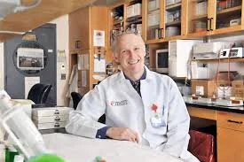
Kenneth Simpson, BVM&S, PhD, Dipl ACVIM, Dipl ECVIM-CA, Professor of Medicine, College of Veterinary Medicine, Cornell University. Dr. Simpson graduated from Edinburgh in 1984 and was awarded a PhD in gastroenterology at the University of Leicester in 1988. He completed an internship at the University of Pennsylvania and a medicine residency at The Ohio State University. He has been a lecturer at the Royal Veterinary College and joined the faculty at Cornell University in 1995. Dr. Simpson is a recipient of the National Phi Zeta and Pfizer awards for research, the BSAVA Bourgelat Award for outstanding contributions to the field of small animal practice and the AVMF/AKC Career Achievement Award in Canine Research. He is past-president of the Comparative Gastroenterology Society.
Dr. Simpson's research interests are centered below the diaphragm, with a focus on inflammatory disease of the GI tract (including the pancreas and liver), host bacterial interactions in health and disease, and culture independent bacteriology.
Canine Biliary disease: Obstruction, Mucocele and Cholecytitis.
Biliary obstruction, cholangitis, mucoceles and more!! An in depth overview of diagnosis and management.
Hepatic Lipidosis: Not just fat cats
This talk will review current thoughts on the etiopathogenesis and management of hepatic lipidosis. It used to be considered”idiopathic” but an underlying disease is present in most cats: another liver disease such as cholangitis, pancreatitis or gastrointestinal disease was present in over 2/3rd of 150 cats in one study.Other common concurrent diseases were neoplasia, urogenital disorders and infectious disease.
Feline Inflammatory Liver Disease: Infectious, Immune or Neoplastic?
Is it infectious, immune or neoplastic? Recent studies have greatly improved our understanding of feline inflammatory liver disease. A systematic approach to diagnosis and treatment will be presented along with new information on bacterial involvement.
Regurgitation What’s the cause?
Regurgitation is the principal sign of esophageal disease. This presentation will review the diagnosis and management of common esophageal disorders.
Hypersalivation: What’s the cause?
Hypersalivation is most commonly associated with disease of the oral cavity, esophagus, stomach and liver. This case based presentation will illustrates how this sign can lead to localizing an underlying cause.
Chronic vomiting: What’s the cause?
A thorough and systematic approach is required to determine the cause of persistent vomiting. An integrated approach based on patient history, physical findings, clinicopathological testing and diagnostic imaging will be presented.
Medical management of gastric disease and persistent vomiting
This presentation will review the treatment of common causes of persistent vomiting such as gastritis, and delayed gastric emptying. This will include “when, why, and how” to use antiulcer, and prokinetic drugs.
Rethinking Canine HGE: Acute Hemorrhagic Diarrhea Syndrome
Sudden onset diarrhea and vomiting accompanied by gastrointestinal hemorrhage (commonly called HHGEE) is a common clinical scenario. Recent studies have linked toxigenic Clostridium perfringens to this syndrome and have critically addressed the impact of treatment on outcome. Are antibiotics needed in every case? Is there a role for probiotics? Why has the syndrome been renamed?
Update on Diagnosis and Management of Granulomatous colitis
Granulomatous colitis (GC) is an inflammatory bowel disease (IBD) defined by infiltration of the colonic mucosa with macrophages. Concurrent cellular infiltrates frequently include neutrophils, lymphocytes and plasma cells. GC is often caused by an underlying infection such as bacteria (e.g., E. coli in Boxers, French Bulldogs), fungi, algae, or protozoa in susceptible dogs. Dogs with GC typically have minimal improvement with empirical treatment (e.g., dietary modification, metronidazole), and it is essential these patients are not immunosuppressed until localized and systemic infections have been ruled out. The prognosis for GC can be very good if a treatable infectious agent is detected. Dogs with GC accompanied by disseminated infections or idiopathic GC have a poorer prognosis.
